


このたびFull textで論文が手に入れられる様になったので、私の医学博士論文を提示し、説明させて頂きます。実は論文は手元にコピーは持っているのですが、頻繁に読む機会があるのではないので、久し振りに全文を読み返してみるのは、楽しみでもあります。
今回は各項目ごとに要訳を添えます。
Scanning Electron Microscopic Study on Double and Single Eyelids in Orientals
アジア人の二重まぶたと一重まぶたの走査式電子顕微鏡での研究;オリエンタルズを東アジア人と訳します。東洋人の蔑視語です。
Kazuhiko Morikawa, M.D., Hiroshi Yamamoto, M.D., Eiju Uchinuma, M.D., and Shohei Yamashina, M.D. Kitasato university, School of Medicine, Sagamihara, Japan
著者 森川一彦:主著者 山本博:共同研究者 内沼栄樹:指導医である北里大学形成外科・美容外科教授 山科正平:技術的指導医である北里大学解剖学教室教授 なお論文発行時点では、前二者は医学博士ではないのでM.D.;医学士の称号だけで、Ph.D.;博士の称号は付けない。教授は当然のごとく医学博士なので称号は付けない。
Abstract.
The conventional theory is that Occidentals have a terminal insertion of the levator aponeurosis at the anterior portion, resulting in a double eyelid, whereas in Orientals this fiber is not present, and therefore results in a single eyelid have been anatomically demonstrated. However, there have been more than a few reports indicating that the anatomical difference between a single eyelid and double eyelid in Orientals cannot be explained by this theory. Therefore, in order to verify the direction of the levator aponeurosis in the eyelids of Orientals, we observed Japanese eyelids using a scanning electron microscope (SEM). As a result of three-dimensional, crosssectional observations using SEM, we were able to confirm the existence of a branch of the levator aponeurosis that runs through the layer of the orbicularis oculi muscle and connects with the levator aponeurosis in the double eyelid, as in the occidental eyelid. This was not seen in the single eyelid. It is thought that this new anatomical finding will become an important fundamental for double eyelid operations in Orientals.
主張したい事はこの抄録に要約されています。抄録は前文ではなく、検索に使うために短文化した論旨です。
従来から、西洋人は挙筋腱膜の終末挿入が、眼瞼の前方にあるために二重まぶたであり、東洋人では、それがないため一重瞼になるというセオリーが提示されていた。しかし東洋人では、このセオリーで説明できない解剖的差異を示す報告がある。そのため、東洋人に置ける挙筋腱膜の走行を同定するため、日本人の眼瞼を走査式電子顕微鏡(SEM)を用いて観察した。3次元的な断面像によって二重瞼では西洋人の様に挙筋腱膜の枝、それも眼輪筋を貫いて繫がっているのを確認した。これは一重瞼では見られなかった。この新しい解剖的知見は、東洋人に置ける重瞼術のために重要な根本的基礎となると考えられる。
Key words: Oriental eyelid—Blepharoplasty—SEM
キーワードは論文検索の際に引かれます。
Blepharoplasty mainly means rhytidectomy in the West, but in Asia it often means a double-eyeli operation in which a single eyelid is made into a double eyelid. When attempting double-eyelid surgery, a fundamental understanding of the anatomical features of the Oriental single eyelid an double eyelid is considered most important. However, although the double eyelid exists also among Orientals, there have been few reports of the anatomy of the double eyelid and the single eyelid in Orientals. The theory that in occidentals the levator aponeurosis runs through the orbicularis oculi muscle layer and attaches to the anterior portion of the eyelid, where it forms a double eyelid, and in Orientals in whom this is not present, a single eyelid forms, was first proposed by Sayoc [1]. This has been supported ever since as Sayoc’s theory. In Japan, Furukawa [2] alone reported that the difference of height at which the levator aponeurosis attaches to the skin determines the factor whether or not there will be a double eyelid or single eyelid, even in Japanese. However, Doxanas and Anderson [3] reported that a single eyelid or double eyelid will occur based on a difference in the height at which the levator aponeurosis and orbital septum fuse. In response to this, reports from Asian countries investigated the differences between the Occidental double eyelid and Oriental single eyelid—differences in thickness of tissue such as skin, subcutaneous tissue, orbicularis oculi muscle, retroorbicularisoculi-fat (ROOF), and pretarsal fat; orbital fat thickness, and the degree of prolapse the existence of pseudoptosis and Mongolian fold; and differences in the size of the tarsus height and orbital cavity, or transverse palpebral fissure. All these factors collectively account for the results [4,5]. Hiraga [6] and Nishiyama et al. [7] explained the single eyelid and double eyelid in terms of a quantitative difference in tissue such as fat, but these findings were not clearly verified numerically. Miyake et al. [8] showed quantitative differences in tissue by MRI imaging, but they could not find the insertion of the Presented at the 15th Congress of the ISAPS, Tokyo, Japan, April 6, 2000 levator aponeurosis to the anterior portion by MRI. It has also been reported that the height of fusion between the orbital septum and levator aponeurosis is equal—a few millimeters above the upper border of tarsus—in all Oriental cases, which is a refutation of the theory by Doxanas and Anderson [3] and others. Similarly, among recent histological studies using light microscopy, there have been some reports in Japan that refute the theory by Sayoc [1], and purport that fibers thought to branch off from the levator aponeurosis and attach to the orbicularis oculi muscle do not reach the anterior portion [9]. In this study, we investigated the continuance of the levator aponeurosis using a SEM in order to clarify the anatomical differences between double eyelids and single eyelids among Japanese.
通常文章では、論旨を解りやすくするために起承転結で記述していく様に求められます。洋語文の論文でもその形を採る事が多いのですが、前文(起)には題がなく他には標題があります。
前文の要旨:先ず私が言いたかったのは、西洋では(アジア人を対象としないという意味)、眼瞼形成術とはしわ、たるみ取り術が主で、アジアでは、重瞼術がしばしば行われること。したがって、この分野の手術はアジア人の解剖構造を知るべきだと考えられる。アジア人にも二重まぶたは存在する。二重まぶたと一重まぶたの解剖の報告は少ない。西洋人では挙筋腱膜が眼輪筋を貫き眼瞼の前葉に付着するのが二重瞼の構造だとされている。日本人でも、二重まぶたでは同様という説がある。引き換えて、二重まぶたと一重まぶたのではほかの要因もあるという説がある。例えば、挙筋腱膜と眼窩隔膜の合流する高さ。皮膚や皮下組織、眼輪筋、ROOF、瞼板前脂肪、眼窩脂肪の厚さなど量的差異。蒙古襞の程度。瞼板高や眼窩の深さの違いなどが重瞼に影響すると報告されてきた。下線部は臨床的にも認められるが、重瞼には影響していない。他の構造的量的差異は臨床的には重瞼と相関しない。また、挙筋腱膜の前葉への挿入はMRIでは見いだせないとの報告もあるが、挙筋腱膜と眼窩隔膜の合流高は皆同じだとし、前者の説を否定している。また最近の光学顕微鏡での研究で、挙筋腱膜が前葉に付着する説を否定するものがある。そこでこの研究では、挙筋腱膜の走行を走査式電子顕微鏡で、日本人における二重まぶたと一重まぶたの解剖構造の差を明らかにするために行います。
Materials and Methods
We studied 10 eyelids from five cadavers (two males, three females) that appeared to the naked eye to be free of facial abnormalities and damage. Age at death ranged from 67 to 92 years. We manually opened the eye, then classified two subjects into a group in which the supra palpebral fold could be seen clearly above the eyelid margin, and three subjects into a group in which the supra palpebral fold could only be seen just above the eyelash near the eyelid margin. We therefore considered the former group to have double eyelids and the latter group to have single eyelids. From each cadaver, we took three sagittal sections 3 to 5 mm in thickness from the central region of the upper eyelid. Of these, one from each cadaver was used as a specimen for light microscopy. The specimens were fixed in Zamboni’s solution, and paraffin migration, whereby 8 mm thin sections were taken and subjected to van Gieson’s stain. The remaining each two samples were prepared in accordance with the tissue-treating method with NaOH in order to observe the fibers of collagen tissue by SEM [10]. The pieces were fixed in 2.5% glutaraldehyde and immersed in a 10% aqueous solution of NaOH for 4 to 7 days at room temperature. The cellular elements were then removed. After being rinsed in distilled water for 1 day, the pieces were placed in 1% tannic acid for 1 to 2 h, rinsed in distilled water, and then postfixed in 1% OsO4 for 2 h. The specimens were dehydrated in a series of grade concentrations of ethanol and t-butyl alcohol, and freeze-dried in a ID-2 dryer. The dried specimens were mounted on a brass stub and coated with gold. Observations were made under an S-4500 SEM by low magnification at ×30 to high magnification at ×100,000. Based on continuous imaging photographed at low magnification connected together, we provided the appearance of continuation of the tissue in a wide visual field. Images that were taken at high magnification confirmed the direction of collagen fibers, and we identified each of the tissues, including the levator aponeurosis.
検体と方法(承):さあ何をするかの説明です。今回の方法のポイントは、光学顕微鏡像と同様に眼瞼の断面を見るのですが、細胞成分を化学的に除去してから見るので、残ったコラーゲン繊維の走行を立体的に追えることです。細かい点は省きますが、上記が定式の方法です。
Results
Light Microscopic Findings :Because the collagen fibers were well visualized by van Gieson’s stain, from the levator aponeurosis descending along the anterior surface of tarsus, branching off in the direction of the orbicularis oculi muscle anteriorly, the collagen fibers were shown to run into the muscle belly of the orbicularis oculi muscle. However, due to the thin sections, the view was interrupted, and we were unable to confirm whether or not the course of the collagen fibers was really continuous (Fig. 1 right side).
Scanning Electron Microscopic Findings: We were able to observe the direction of the levator aponeurosis, continuous with the levator palpebrae superioris muscle, using the SEM because it allows observation of cross-sections three dimensionally. It was easy to identify the levator palpebrae superioris muscle and Mu¨ller’s muscle posterosuperior to the conjunctival fornix by observing the directions of the bundle. By following the direction of these muscles towards the eyelid margin, we were able to detect each of the layers: the tarsus, aponeurosis, fat, and orbicularis oculi muscle (Fig. 1 left side). Furthermore, in order to identify the levator aponeurosis, we scanned and observed in the direction of the eyelid margin from the height of the upper margin of tarsus using high magnification. In samples of double eyelids obtained from cadavers, the levator aponeurosis was observed as a bundle of dense collagen fibers running in the same direction at the height of the upper margin of tarsus; it did not make contact with the tarsus until one-third of the height of the tarsus (Fig. 2). While this levator aponeurosis further ran inferiorly along the anterior surface of tarsus, a large number of collagen fiber branches could be seen running into the muscle belly of the orbicularis oculi muscle positioned anteriorly. In addition, the position in which the collagen fibers branch off was inferior to the orbital septum even at the highest position. These fibers were shown also by high magnification to be continuous with the levator aponeurosis anterior to the tarsus. High magnification also showed the branches to appear dense. Although there was slight disarray, the direction of the fibers was uniform, and similar to the fibers of the aponeurosis above (Fig. 3). Observation of the character and unidirection of the fibers suggested that the strength to open the eyelid is transmitted to the anterior portion similarly to the upper levator aponeurosis. This bundle of collagen fibers penetrates the orbicularis oculi muscle layer and inserts at the subcutaneous layer whereby it appears wavelike. This connects to collagen fibers that are not uniform in direction and are composed of subcutaneous connective tissue, but it does not reach the dermis (Fig. 4). In single eyelids of cadavers, the direction of the levator aponeurosis anterior to the tarsus was the same as in double eyelids. But the difference with a double eyelid was that anterior branches at any position were out of view, and connected with subcutaneous tissue of the eyelid margin (Fig. 5).
結果(転)はこうなりましたという事ですが、まだ何も主張していません。長文になりますが要約すると:二重まぶたでは、上眼瞼挙筋腱膜から密なコラーゲン繊維が前方に向かって枝分かれして走行し、眼窩脂肪の下を通って眼輪筋を貫いて、皮下組織に繋がっている。一重まぶたでは瞼縁まで枝分れがない。判ったことはこれだけです。
Discussion and Conclusion
In this study we used a scanning electron microscope to study the anatomy of the eyelid; it was the first time this method was used. Although we could not study quantitative differences in skin, subcutaneous tissue, orbicularis oculi muscle, and fat, nor the positioning of fusion between the orbital septum and levator aponeurosis from the findings of the SEM, we were able to view three dimensional images of the eyelid. As a result, we were able to clarify the continuity of branch fibers of the levator aponeurosis. When we investigated the double eyelid and single eyelid among Japanese, we found similarities with the findings of Sayoc [1] regarding the double eyelid. That is, the bundle of collagen fibers that branch off from the levator aponeurosis run through the orbicularis oculi muscle layer and insert at the subcutaneous tissue layer. However, in contrast to Sayoc’s findings, we found that this terminal fiber does not directly contact the skin, but is continuous with the collagen fibers in subcutaneous tissue. It is thought that a double eyelid will result sufficiently even if this terminal fiber does not reach the dermis. In single eyelids, we observed that the levator aponeurosis runs along the tarsus inferiorly without branching off until the eyelid margin, and reaches the subcutaneous tissue of the eyelid margin. This result concerning the single eyelid was similar to previous reports. Whether or not the levator aponeurosis branches off below the orbital septum, passes through the orbicular is oculi muscle layer, and runs continuous with subcutaneous tissue is considered the determining factor for a double eyelid or a single eyelid (Fig. 6). Recently in Japan, there has been a report in which the authors could not find the insertion of the levator aponeurosis at the anterior portion by MRI [8], and a report in which fibers thought to be attached to the orbicularis oculi muscle were not continuous with the levator aponeurosis and did not extend to the dermis [9]. Sayoc’s theory, which explains the anatomical difference in eyelids between Occidentals and Orientals, has not been able to account for anatomical difference between double eyelids and single eyelids in Orientals. However, because a SEM, which we used, was more useful than other methods of investigation, it was possible to obtain clear results of the direction of the levator aponeurosis. We found that an important factor accounting for the anatomical difference between single eyelids and double eyelids in Orientals is the anatomical difference between the eyelids in Occidentals and Orientals reported by Sayoc. Oriental aesthetic surgeons have many clinical opportunities to perform double eyelid operations to transform an Oriental single eyelid into a natural-looking Oriental double eyelid. In view of the results of the present histological study, we think that if double eyelid surgery is to be performed, it would be adequate if transmission of levator muscle force towards the anterior portion could be obtained by connecting a part of the levator aponeurosis with the most superficial layer of orbicularis oculi muscle, or by connecting a part of the levator aponeurosis with the most superficial layer of orbicularis oculi muscle and the subcutaneous tissue.
考察と結語(結)です。:要約することで論旨を説明します。西洋人は全員二重まぶたなのに、東洋人では二重まぶたと一重まぶたが混在している。両者は解剖構造の何が違うのかについて日本でもいくつか報告されていて、その中の一つには挙筋腱膜からの皮膚への連続性は認めなかったというものもある。しかし今回の走査電子顕微鏡による三次元的画像解析により、挙筋腱膜からの枝分れ繊維を描出できた。
結論としては:東洋の美容外科・形成外科医は、東洋人の一重まぶたを、自然に見える東洋人の二重瞼に改良する重瞼術を施行する機会が多い。ここに提示した組織学的研究の結果からの観点からして、重瞼術が施行されるならば、挙筋の一部が眼輪筋の最浅層か皮下組織に連結するによって、挙筋力が眼瞼前葉に向かって伝達されるようにすることが適切であると考える。つまり、繋がりを作ることが唯一、重瞼術の必要十分条件だといえる。
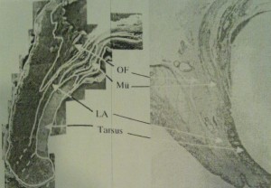
Fig. 1. (Right) Light microscopic view. It seems that the levator aponeurosis branches anteriorly into the orbicularis oculi muscle (van Gieson’s stain, original magnification ×1). (Left) SEM view. White lines show each layer of the sagittal section of the eyelid (original magnification ×50). Mu¨ ; Mu¨ller’s muscle; LA 4; levator aponeurosis; OF ; orbital fat.
Fig. 2. (Upper) SEM view at the level of the upper one-third portion of tarsus of a double eyelid. White lines show each layer of eyelid. The levator aponeurosis runs down the anterior surface of the tarsus, and it has multiple lamella (original magnification ×50). T; Tarsus; Post; postaponeurotic fat. (Lower) A highly magnified view of the levator aponeurosis. This is formed by bundles of collagen fibers which run in the same direction and are dense (original magnification ×10,000).
Fig. 3. (Upper) SEM view of the middle one-third portion of tarsus of the double eyelid. This shows the branch fibers of the levator aponeurosis that run between the bundles of fibers of the orbicularis oculi muscle. The fibers of the levator aponeurosis connecting the branch fibers (original magnification ×60). M ; orbicularis oculi muscle. (Lower) SEM highly magnified view of the branch fiber. The fiber is the same as in Figure 2 (original magnification ×10,000).
Fig. 4. SEM view of the eyelid margin. The schema shows that the branch fibers insert into the subcutaneous tissue. The supra palpebral fold is beneath this fiber (original magnification ×60).
Fig. 5. (Upper) SEM view at the upper half portion of the tarsus of a single eyelid. The levator aponeurosis does not branch, and runs down for the eyelid margin (original magnification ×30). (Lower) SEM view of the lower half of the tarsus of a single eyelid. The levator aponeurosis inserts at the subcutaneous tissue and pretarsal connective tissue (original magnification ×30).
Fig. 6. Anatomic difference between a double eyelid (left) and a single eyelid (right) in an Oriental. The fundamental difference is whether or not there are branch fibers. Mu¨ ; Mu¨ller’s muscle;ROOF ; retro-orbicularis-oculi-fat; OF ; orbital fat;LA ; levator aponeurosis; M ; orbicular is oculi muscle.
図の説明は載りました。実はまだ、このサイト内では論文から採った画像を転載できる機能を把握していないので、その点は残念ですが、これから努力してみます。画像が結構面白いんんですよ!
References
1. Sayoc BT: Absence of superior palpebral fold in slit eyes: An anatomic and physiological explanation. Am J Ophthalmol 42:298, 1956
2. Furukawa M: Aesthetic surgery of Oriental eyelids. Aesth Plast Surg 1:139, 1977
3. Doxanas MT, Anderson RT: Oriental eyelids. An anatomic study. Arch Ophthal 102:1232, 1984
4. Siegel R: Surgical anatomy of the upper eyelid fascia. Ann Plast Surg 13:263, 1984
5. Song RY, Song YG: Double eyelid operations. Aesth Plast Surg 9:173, 1985
6. Hiraga Y: The double eyelid operation and augmentation rhinoplasty in the Oriental patient. Clin Plast Surg 7:553, 1980
7. Nishiyama S, Ueno F, Hiraga Y, Yamahata A: Consideration on the mechanism of double eyelid formation asymmetry of the width in the double eyelid operation (Japanese). J Jpn Soc Aesth Plast Surg 11:67, 1989
8. Miyake I, Tange I, Hiraga Y: MRI findings of the upper eyelid and their relationship with single-and double-eyelid formation. Aesth Plast Surg 18:183, 1994
9. Tsurukiri K: Anatomy of upper lid (histological examination of a sagittal section through the upper lid-first report) (Japanese). Jpn J Plast Reconstr Surg 14:137, 1992
10. Ohtani O: Three-dimensional organization of the connective tissue fibers of the human pancreas: A scanning electron microscope study of NaOH-treated tissues. Arch Histol Jpn 50:557, 1987
ここには、参考文献が羅列されていますが、単に羅列でなく、論文中でのこれまでの議論や、方法論を参照するために、文中に括弧数字で引用した事が示してあるものだけを載せています。 論文中に引用すれば著作権を侵さないのですが、逆に論文とその本や雑誌は、国際的にチェックされ検索サイトにも登録されます。
論文が載っている著作物の読者数に応じて、引用された論文の読者数が計算されて、引用数の多寡が、その論文の科学的価値の基準とされます。これをインパクトファクター(IF)といいます。論文が他の科学者に引用されるという事は、相手がその論文の有用性、ひいてはその価値を認めていると考えるからです。
したがって、IFがその科学者の作製した論文の質と量を示し、教室や講座等の集団内のIFの和が、研究費や員数の配分に直結し、大学や研究機関単位の総和もその評価につながり、結果といて公的な補助金の配分に繫がるのです。
もっとも、私はこの論文を作製するに当たっては、十編の論文しか参照にしていない訳ではありません。ざっと百編以上は読み下しました。
そりゃそうです。Pubmedで上眼瞼: upper eyelid で検索すると5000編以上、二重瞼: double eyelid では1000編以上、しぼって挙筋腱膜: Levator aponeurosis でも380編もの論文がヒットするのです。先ずこの辺りを検索しました。逆に考えると、その中からこの論文を書く為に参考になる、有用性のある論文を探し出すのに苦労したのです。結果よく見ると、十編の参考文献のうち半分の5編に対しては私は反論しています。しかしそれだけに、私の論文の画像による反証は科学的価値が高いといえると思います。もしこの論文の、keywordでPubmedを検索すると、Oriental(Asian) eyelid では312編、blepharoplasty では2798編、SEM(scannig electron microscope) では119748編も見つかりますが、前2者では159編、それもアジア人ばかりです。3つとも入れると、当然この論文しか出ません。
Correspondence to Dr. Kazuhiko Morikawa, Department of Plastic and Reconstructive Surgery, Kitasato University School of Medicine, 1-15-1 kitasato, Sagamihara, Kanagawa 228- 8555, Japan. Aesth. Plast. Surg. 25:20–24, 2001
連絡先は、他の科学者からの反論や、参照時のコンタクトのために必要なのですが、連絡はありませんでした。他の形成外科医や美容外科医は興味がないのでしょうか?、それとも二重瞼と一重瞼の問題は世界人類の一部にしか関係ないからでしょうか?。 実は論文の掲載誌のIFが低いからです。私はJSAPSに参加している日本の美容外科医約1000人のうちの200人とは毎年学会での情報交換していますが、この論文を読んでくれたのはそのうち10人に一人もいませんでした。重瞼術を行うのに興味ないなんて、なんて我が国の美容外科医は非科学的、ビジネス的なのかと、遺憾に思います。
DOI: 10.1007/s002660010088 © 2001 Springer-Verlag New York Inc.
これが、この論文の掲載誌の発行社です。多くの医学雑誌を刊行しているので、美容医療のAesthetic Plastic surgery 誌は、サイトの端っこにしかありませんでした。
という訳で、今回美容外科医にとってとても為になるはずの論文である、二重瞼と一重瞼の違いの研究を要説しました。科学的論拠に則って美容医療をしようじゃありませんか?。糸で繋ぐだけでも切開するにしても、この議論の要旨は頭に入れておきましょう。
日本国内の医学博士論文は、国会図書館に提出されます。ですから、インターネットでも閲覧できます。ちなみに検索してみると、美容外科分野(形成外科分野でない。)の研究で医学博士の称号を得た医師は、他に一人もいませんでした。生涯一美容外科医を目指す私が唯一です。天命と捉えます。もちろん、美容整形医の走りである父も生前、大変誇りにしていたようです。ただしその頃から、私が医学的知識を語ると、父は「よく研究されていますねえ。教授!」と揶揄する様になりました。美容医療はそんな斯界なんです。
次回のお勉強シリーズは、リフト系を載せたいと思います。その前に論文から画像が載せられたら、それも。